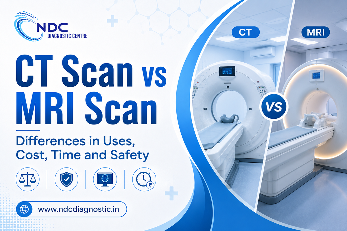
CT and MRI can both produce detailed images from inside the body, but they work in very different ways and are not interchangeable.
A CT scan uses X-rays and computer processing to create cross-sectional images. MRI uses a strong magnetic field and radiofrequency energy and does not use ionising X-rays. CT is particularly useful when imaging needs to be obtained quickly, including many emergency situations. MRI often provides more detailed contrast between different soft tissues, which can be useful for the brain, spinal cord, joints, muscles and other structures.
Neither test is automatically better. The useful question is not "Which machine is more advanced?" but "Which scan gives the information needed for this particular problem?"
|
Feature |
CT Scan |
MRI Scan |
|
How images are created |
X-rays and computer reconstruction |
Strong magnetic field and radiofrequency energy |
|
Ionising radiation |
Yes |
No |
|
Scan speed |
Generally faster |
Usually takes longer |
|
Often useful for |
Trauma, lungs, many fractures, acute bleeding and rapid assessment |
Brain, spinal cord, joints, ligaments, muscles and many soft tissues |
|
Bone detail |
Often very good |
Can assess bone marrow and surrounding soft tissues |
|
Soft-tissue contrast |
Good for many conditions |
Often superior for selected soft tissues |
|
Noise |
Usually limited |
Often produces loud knocking or tapping sounds |
|
Movement |
Shorter scan can make motion easier to manage |
Staying still for longer is important |
|
Contrast when required |
Commonly iodinated contrast |
Commonly gadolinium-based contrast |
|
Implants and devices |
Fewer magnetic-field restrictions |
Requires MRI safety screening |
|
Emergency use |
Frequently useful because image acquisition is fast |
Useful in selected emergencies but typically slower |
|
Cost |
Depends heavily on body area and protocol |
Often more expensive for comparable studies, but not always |
|
Main practical limitation |
Uses ionising radiation |
Longer scan and magnetic-field safety considerations |
The comparison is only a guide. A CT chest, MRI knee and CT angiography are very different examinations, so the modality name alone does not tell you which scan is appropriate.
CT stands for computed tomography.
During CT imaging, an X-ray source rotates around the body and collects information from multiple angles. A computer reconstructs that information into cross-sectional images, often described as slices. CT can show bone, organs, blood vessels and soft tissues in considerable detail.
MRI stands for magnetic resonance imaging.
Instead of X-rays, an MRI scanner uses a powerful magnetic field, radiofrequency energy and a computer to create images. Signals are generated mainly from hydrogen protons in tissues containing water and fat.
This difference is important because MRI is not simply a more advanced version of CT. Each technology highlights different anatomical features and comes with different practical and safety considerations.
The reason for ordering one scan rather than the other often becomes clearer when you look at what the doctor needs to assess.
CT is widely used for rapid evaluation of trauma and acute illness.
Examples include assessment of:
One of CT's biggest practical advantages is speed. In an emergency, obtaining images quickly can matter as much as the level of anatomical detail.
The FDA notes that CT is used to diagnose disease, trauma and abnormalities and can also guide treatment and procedures.
MRI is often particularly useful when the clinical question involves soft tissues.
This can include:
The FDA notes that MRI generally provides better soft-tissue contrast than CT and can distinguish between tissues such as fat, water and muscle more clearly in many situations.
That does not mean MRI is more accurate for every problem. Accuracy depends on what the scan is being asked to show.
Patients often search for the "better" scan for a particular body part, but even within the same area, the preferred test can change depending on the suspected condition.
Both CT and MRI have important roles in brain imaging.
CT is commonly valuable when a rapid assessment is needed, including certain head injuries and acute neurological emergencies.
MRI can provide more detailed soft-tissue information for many neurological conditions because of its high contrast between different types of brain tissue.
The choice may therefore depend as much on urgency and the suspected problem as on image detail.
MRI is often useful for evaluating the spinal cord, discs, nerves and surrounding soft tissues.
CT can provide excellent detail of the vertebral bones and can be particularly useful for certain fractures or when MRI cannot be performed.
CT is very useful for visualising bone anatomy and complex fractures.
MRI can provide additional information when the concern extends beyond the bone itself, such as bone marrow changes, cartilage, tendons, ligaments or nearby soft-tissue injury.
CT has an important role in imaging the lungs and many structures within the chest because it can capture detailed images quickly.
MRI can also be used for selected chest and cardiovascular questions, so saying that MRI "cannot scan the chest" would be incorrect.
MRI is frequently chosen when doctors need a detailed view of structures such as ligaments, tendons, cartilage and muscles.
For example, a knee injury may involve bone, ligament, cartilage or several structures at once. The most appropriate examination depends on what is suspected.
CT image acquisition is generally much faster than MRI.
A CT scan itself may be completed rapidly, although preparation, contrast administration and positioning can increase the total appointment time.
MRI usually takes longer because several imaging sequences may need to be collected. FDA guidance notes that a typical MRI examination can take approximately 20 to 90 minutes depending on the body part and type of scan.
That does not mean every MRI takes an hour or every CT takes only a few minutes.
The total time can change depending on:
This distinction matters when comparing online claims about scan time. The time spent collecting images and the total time at the diagnostic centre are not necessarily the same.
There is no meaningful single price for "a CT scan" or "an MRI scan."
Current Mumbai listings in September 2026 show why. Public CT prices vary according to the procedure, with examples such as a contrast-enhanced CT face starting around ₹4,125 and several CT angiography studies listed around ₹5,520. MRI listings also vary considerably, from approximately ₹3,152 for a left knee MRI to ₹8,970 for a plain cardiac MRI, while more extensive MRI studies can cost substantially more.
These examples are not like-for-like comparisons. They illustrate why comparing a CT of one body region with an MRI of another gives a misleading picture of cost.
Pricing can depend on:
MRI often costs more than a comparable routine CT study, but that should not be treated as a universal rule.
Before booking, confirm the exact investigation written on the prescription and what the quoted price includes.
The biggest safety difference most patients notice is radiation.
CT uses X-rays, which means it involves ionising radiation. MRI does not use ionising radiation.
That fact should not be simplified into "MRI is safe and CT is dangerous."
The FDA states that when CT is used appropriately, its diagnostic benefits far exceed the risks. Radiation exposure is kept as low as reasonably practical for the examination, and the amount varies according to the type of CT, body area, patient and scanning technique.
Children are more sensitive to ionising radiation than adults, which is one reason CT techniques are adjusted according to age and size where appropriate.
A medically necessary CT should not be avoided simply because it uses radiation. The clinical value of getting the required diagnosis needs to be considered alongside the exposure.
MRI avoids ionising radiation but has a different set of safety considerations because of its powerful magnetic field.
Having metal or an implanted medical device does not automatically mean you cannot undergo MRI.
MRI devices and implants may be classified according to their safety in the MR environment. Some are MR Safe, some are MR Conditional and some are MR Unsafe. An MR Conditional device may be scanned only when the conditions specified for that device are met.
Before MRI, tell the imaging team about any:
The exact device should be identified so its MRI safety information can be checked.
The FDA advises that patients with implanted devices should not undergo MRI unless the device has been positively identified as MR Safe or MR Conditional under appropriate conditions.
MRI can also feel uncomfortable for people with significant claustrophobia. The scanner is noisy, staying still is important and some examinations take considerably longer than CT.
If you have severe claustrophobia or anxiety about the scan, discuss it with the healthcare team before the appointment so the examination can be planned appropriately.
Contrast is used in some CT and MRI examinations to make particular tissues, organs or blood vessels easier to see.
Not every CT or MRI requires contrast.
CT commonly uses iodinated contrast material when enhancement is needed.
MRI commonly uses gadolinium-based contrast agents. These are different from the iodinated contrast agents commonly used in CT.
Before receiving contrast, tell the imaging team about relevant medical information, including:
Contrast should not automatically be described as dangerous for people with kidney problems. The type of contrast, kidney function and clinical need all matter.
Patients should also not stop prescribed medicines or refuse necessary contrast based only on information found online. Follow the instructions given for the specific examination.
Pregnancy is another area where overly simple advice can be misleading.
MRI does not use ionising radiation and is often useful when imaging is clinically necessary during pregnancy. Current guidance does not show proven harm from appropriately performed non-contrast MRI, although the examination should still be done for a valid medical reason.
CT uses ionising radiation, but that does not mean it can never be performed during pregnancy.
RadiologyInfo notes that when CT is medically necessary and the required information cannot be obtained adequately another way, the examination may still be the appropriate option. The decision depends on the body area, urgency and clinical question.
Contrast decisions require separate consideration. Gadolinium contrast for MRI is generally limited during pregnancy and used only when its additional diagnostic value is important.
If you are pregnant or think you may be pregnant, tell the imaging team before either a CT or MRI.
Neither scan is universally better.
CT may be preferred when:
MRI may be preferred when:
The right scan depends on the body part, suspected condition, urgency, previous imaging, implant safety, contrast requirements and other individual factors.
Do not replace a prescribed CT with an MRI simply because MRI does not use ionising radiation. They do not provide identical information.
You lie on a table that moves through a relatively short, ring-shaped scanner.
The machine acquires images as you pass through the opening. For some examinations, you may be asked to briefly hold your breath.
Contrast may be given by injection or another route depending on the study.
Image acquisition is usually fast, although the total appointment can take longer when preparation or contrast is required.
You lie on a table that moves into the MRI scanner.
MRI examinations involve repeated imaging sequences and produce loud knocking or tapping sounds. Ear protection is commonly provided.
You need to remain still because movement can reduce image quality.
Depending on the scan, contrast may be given intravenously.
Preparation varies, so do not assume every CT or MRI requires fasting.
Before the appointment:
Do not change medicines unless your healthcare professional specifically tells you to do so.
NDC Diagnostic Centre's current radiology page includes CT scans and MRI imaging among its radiology services. The same page also describes X-ray, ultrasound, mammography and bone densitometry services.
You can review NDC's radiology services for imaging information.
Because the exact examination and availability can vary by location, check the NDC Diagnostic Centre locations and confirm the prescribed scan before travelling.
NDC's appointment page also includes CT Scan and MRI among the available service selections.
If your doctor has prescribed CT or MRI, confirm the exact body area, whether contrast is required, preparation instructions, current price and availability before booking.
CT uses X-rays to create cross-sectional images, while MRI uses a strong magnetic field and radiofrequency energy. CT is generally faster, while MRI often provides greater detail for many soft tissues.
Neither is better for every condition. CT can be more useful for rapid emergency imaging, lungs, many fractures and acute bleeding. MRI can be particularly useful for the brain, spinal cord, joints, ligaments and other soft tissues.
MRI does not use ionising radiation, while CT does. However, MRI has magnetic-field safety considerations involving certain implants and devices. A medically necessary CT should not be considered unsafe simply because it uses radiation.
MRI does not use ionising radiation. It creates images using strong magnetic fields and radiofrequency energy.
CT image acquisition is generally faster. MRI usually takes longer because multiple imaging sequences may be required and the patient needs to remain still.
Not in general. MRI may show certain soft tissues more clearly, while CT may be more useful for other clinical questions. Accuracy depends on what the doctor is trying to assess.
Possibly. Many implants are MR Conditional rather than automatically unsafe. The specific implant or device must be identified and checked against its MRI safety conditions before the scan.
MRI often costs more than a comparable routine CT examination, but this is not universal. Cost depends on the body area, contrast, imaging protocol and facility. Compare prices for the exact prescribed examination rather than comparing CT and MRI as broad categories.
No. CT commonly uses iodinated contrast, while MRI commonly uses gadolinium-based contrast agents when enhancement is needed. Not every CT or MRI requires contrast.