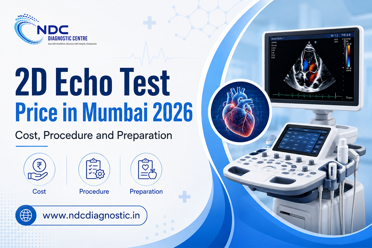
If your doctor has advised a 2D Echo, cost is often one of the first practical questions. Current online listings in Mumbai show that echocardiography prices vary by diagnostic centre, location and the exact type of Echo prescribed. Tata 1mg currently lists an Echo in Mumbai starting at ₹1,380, while another Mumbai pricing source places the broader market range around ₹2,000 to ₹4,000.
The cheapest advertised price is not always directly comparable with another centre's quote. One provider may be showing a promotional starting rate, while another may include Colour Doppler, reporting or a different type of echocardiography.
If you are planning a 2D Echo at NDC Diagnostic Centre, confirm the current price and availability with your preferred centre before booking.
A practical current market estimate for an Echo in Mumbai is approximately ₹1,400 to ₹4,000. The final amount depends on the centre, location, type of echocardiogram and what is included in the quoted price.
|
Price factor |
Why it matters |
|
Type of Echo |
A routine transthoracic Echo differs from stress and transesophageal echocardiography |
|
Colour Doppler |
Some centres include Doppler assessment while others describe or price it separately |
|
Diagnostic centre |
Charges can vary by provider and location |
|
Report and interpretation |
Check whether reporting is included in the quoted amount |
|
Consultation |
A cardiology consultation may be charged separately |
When asking for the price, mention exactly what is written on your prescription. If it says "2D Echo with Colour Doppler", asking only for an "Echo price" may not give you an accurate comparison.
A standard Echo study commonly involves ultrasound imaging of the heart, measurements of the chambers and valves, assessment of heart muscle movement and evaluation of pumping function.
Doppler measurements may also be performed when required to assess blood flow through the heart and valves.
Before comparing prices between two centres, ask whether the quote includes the complete scan, Doppler assessment if prescribed, interpretation and report.
A 2D Echo, or two-dimensional echocardiogram, is an ultrasound examination of the heart. It uses sound waves to create moving images while your heart is beating.
These images allow healthcare professionals to assess the size and shape of the heart, how the heart muscle moves and pumps blood, and how the valves are working. Doppler echocardiography can also provide information about blood flow through the valves.
A doctor may recommend an Echo when more information is needed about:
An Echo does not provide the same information as an ECG. An ECG records the heart's electrical activity, while an Echo shows its structure and movement.
NDC Diagnostic Centre lists ECG, 2D Echo, computerised stress testing, BP monitoring and Holter monitoring among its cardiology services.
A standard transthoracic Echo is performed from outside the chest.
You will usually need to uncover the upper part of your chest and may be given a gown. You then lie on an examination couch, commonly on your left side.
Gel is applied to the chest, and an ultrasound probe is moved over different areas to capture images of the heart from several angles. You may be asked to change position or briefly adjust your breathing while certain images are being recorded.
ECG electrodes may also be attached to the chest to track the heartbeat during the scan.
You may feel some pressure from the probe when the healthcare professional needs a clearer image, but a standard transthoracic Echo is a non-invasive examination.
It uses ultrasound rather than ionising radiation.
Once the required images and measurements have been collected, the gel is removed and the test is complete.
For a standard transthoracic 2D Echo, fasting is generally not required.
Mayo Clinic states that people can usually eat and drink normally before a standard transthoracic echocardiogram and that most Echo examinations require little special preparation.
Preparation changes for some other types of Echo.
A transesophageal echocardiogram may require several hours without food or drink because the ultrasound probe is passed into the oesophagus. Certain stress Echo procedures can also have specific preparation instructions, including restrictions related to caffeine or medicines.
This distinction also matters if you are having an Echo as part of a full health check-up.
Some NDC health check-up packages include 2D Echo along with fasting blood investigations. Those packages instruct patients to fast for 10 to 12 hours because several blood tests are included.
That package instruction should not be treated as a fasting requirement for every standalone standard 2D Echo.
If several investigations are scheduled during the same appointment, follow the preparation instructions given for the complete set of tests.
A routine transthoracic Echo generally needs very little preparation.
Wear clothing that allows convenient access to the chest and follow any instructions provided when the appointment is booked.
It can be useful to carry:
Do not stop or change prescribed medicines on your own before the test.
If a medicine needs to be adjusted for a particular cardiac investigation, follow the instructions given by your treating healthcare professional.
If your prescription mentions a stress Echo, transesophageal Echo or another specialised form of echocardiography, ask for preparation instructions for that specific test.
A standard echocardiogram commonly takes around 30 to 40 minutes.
The exact duration can vary according to the images and measurements needed and whether additional Doppler assessment is required.
The scan duration and report delivery time are separate. If you need the report for an upcoming consultation, confirm the expected reporting time with the diagnostic centre when you book the test.
An Echo report contains several measurements that describe different aspects of heart structure and function.
These findings are interpreted together. One measurement alone does not usually provide a complete picture of heart health.
Ejection fraction, often abbreviated as EF, describes how much blood the left ventricle pumps out when it contracts.
It is one measurement used to understand heart pumping function, but it needs to be considered alongside other Echo findings and clinical information.
An echocardiogram measures the dimensions of different heart chambers.
These measurements can provide useful information when a doctor is assessing conditions involving the heart muscle, blood pressure, valves or other structural changes.
The Echo shows how the valves open and close.
Doppler assessment can provide information about whether a valve has become too narrow, known as stenosis, or whether blood is leaking backwards through it, known as regurgitation.
An Echo allows clinicians to observe how different parts of the heart muscle move during each heartbeat.
If one area moves differently from expected, the finding needs to be interpreted together with the rest of the Echo, symptoms and other cardiac investigations.
Doppler echocardiography evaluates how blood moves through the heart and valves.
It can provide useful information about valve function and certain pressure differences.
If your report contains unfamiliar terminology, discuss it with the doctor who requested the Echo rather than trying to interpret individual values separately.
An ECG and a 2D Echo both assess the heart, but they answer different questions.
An ECG records electrical activity and provides information about heart rate, rhythm and electrical conduction.
A 2D Echo uses ultrasound to show physical structures such as the chambers, heart muscle and valves, along with pumping function.
This explains why a doctor may recommend both tests. One does not automatically replace the other.
For a more detailed explanation, read NDC's guide on ECG vs 2D Echo vs TMT.
A standard resting 2D Echo does not directly show narrowing inside the coronary arteries.
Its main purpose is to assess the structure and function of the heart.
It can show how the heart muscle moves, how effectively the heart pumps and whether there are abnormalities involving the valves or chambers. A routine transthoracic Echo, however, does not directly tell you the exact location or percentage of a narrowing inside a coronary artery.
This means a normal standard Echo should not be interpreted as proof that the coronary arteries are completely free of disease.
If coronary artery disease is suspected, the doctor decides whether another investigation is required based on symptoms, risk factors, physical examination and previous test results.
NDC Diagnostic Centre provides a range of cardiac investigations, including ECG, 2D Echo, computerised stress testing, BP monitoring and Holter monitoring.
You can check the NDC Diagnostic Centre locations when looking for a centre in Mumbai, Navi Mumbai or Thane. NDC's location directory currently groups centres across these areas.
Because services can differ by location, confirm that the exact cardiac test on your prescription is available before travelling.
For patients around Kalyan, the NDC Diagnostic Centre in Kalyan specifically lists 2D Echo, Colour Doppler, ECG and TMT among its services.
When comparing centres, check:
The lowest displayed online price is not always the most useful comparison. The exact test and what is included matter as much as the starting amount.
NDC's cardiology services include 2D Echo along with ECG, computerised stress testing, BP monitoring and Holter monitoring.
If your doctor has advised an Echo, you can review NDC's cardiology services and confirm the current test price and availability with your preferred centre before booking.
Selected NDC health check-up packages also include 2D Echo together with multiple other investigations. Package pricing should therefore be considered separately from the cost of a standalone Echo.
Current online listings suggest that an Echo in Mumbai can cost roughly ₹1,400 to ₹4,000 depending on the provider, type of study and what is included. Tata 1mg currently shows a starting price of ₹1,380.
Fasting is generally not required for a standard transthoracic 2D Echo. You can usually eat and drink normally unless your doctor or diagnostic centre has given different instructions for your specific test.
Yes. People can generally eat normally before a routine transthoracic Echo. If you are also having fasting blood tests or a health package during the same visit, follow the preparation instructions for those investigations.
A standard echocardiogram commonly takes around 30 to 40 minutes, although the exact time varies according to the study and measurements required.
A standard transthoracic Echo is non-invasive and is generally well tolerated. You may feel some pressure from the ultrasound probe against the chest while particular images are being obtained.
A standard resting Echo does not directly show the exact location or percentage of coronary artery narrowing. Its main role is to evaluate heart structure, valves, wall movement and pumping function.
An ECG records the heart's electrical activity, including rate and rhythm. A 2D Echo uses ultrasound to assess the heart's physical structure and function.
No. A standard transthoracic echocardiogram uses ultrasound sound waves and does not use ionising radiation.